


 February 02, 2026 - BY Admin
Understanding DICOM Images: The Backbone of Digital Radiology Reporting
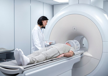
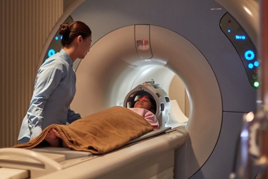
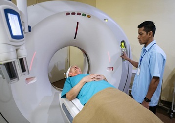
In the world of modern medicine, an MRI or CT scan is much more than just a "picture." It is a complex data set that contains life-saving information.
Behind every high-resolution scan you see on a monitor lies a global standard called DICOM (Digital Imaging and Communications in Medicine).
As a radiologist, I often explain to my clinical colleagues and tech partners that without DICOM, the seamless exchange of medical images across the globe would be impossible. At AITeleRadiology, DICOM is the universal language that allows us to receive a scan from a remote village in India and provide an expert report within minutes.
This 2000-word deep-dive explores the architecture of DICOM, its role in teleradiology, and why it remains the undisputed backbone of digital healthcare in 2026.
1. What exactly is DICOM? More Than Just a File Format
Most people are familiar with JPEG or PNG files for photos. However, in medical imaging, these formats are insufficient. A JPEG file only stores pixel data. A DICOM file, on the other hand, is a "Container" that holds two distinct layers of information:

A. The Image Data (Pixels)
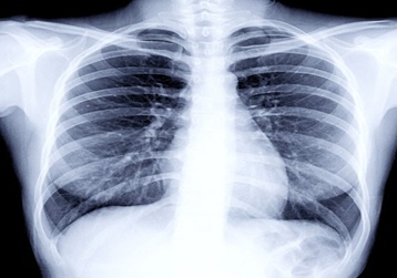
This is the visual representation of the body part. Unlike compressed web images, DICOM stores images in High Bit-Depth (often 12-bit or 16-bit). This allows radiologists to adjust the "Window" and "Level," revealing subtle details in bone or soft tissue that are invisible to the naked eye.
B. The Metadata (The Header)
Every DICOM file has a "Header" that contains:
- Patient Demographics: Name, Age, Sex, and Patient ID.
- Modality Details: Was it a 3T MRI or a 64-slice CT? What were the KVp and mAs settings?
- Spatial Orientation: Exactly where in 3D space the slice was taken (essential for surgical planning).
2. Why Can’t We Just Use WhatsApp or Email for Scans?
A common question from smaller clinics is: "Can I just take a photo of the film and WhatsApp it to you?" From a clinical and legal standpoint, the answer is a firm No.
The Loss of Diagnostic Integrity
When you convert a DICOM image to a JPEG or a photo, you lose "Metadata" and "Dynamic Range." A radiologist cannot "zoom in" or "re-window" a photo to see a tiny tumor hidden in the shadows. DICOM ensures Lossless Compression, preserving every single bit of diagnostic data.
Compliance and Security
DICOM is designed to meet HIPAA (Health Insurance Portability and Accountability Act) and Indian healthcare data laws. It allows for encrypted transmission, ensuring that a patient’s sensitive medical data doesn't end up in the wrong hands.
3. The Technical Pillars: PACS, RIS, and DICOM
To understand how AITeleRadiology works, you must understand the ecosystem where DICOM lives.
- Modality: The MRI or CT machine that generates the DICOM data.
- PACS (Picture Archiving and Communication System): Think of this as the "Digital Library" or hard drive where DICOM files are stored.
- RIS (Radiology Information System): The software that manages patient scheduling and billing.
- DICOM Viewer: The specialized workstation software that radiologists use to manipulate and read the images.
4. How DICOM Fuels Teleradiology in India
The true magic of DICOM is Interoperability. It means a GE machine in a small town can "talk" to a Siemens PACS system in a metro city, which then displays the image on a Barco medical monitor for a specialist.
[Image of teleradiology workflow diagram]
At aiteleradiology.in, our workflow is built on this precision:
- Step 1: The local technician pushes the DICOM study to our cloud server.
- Step 2: Our system automatically extracts the metadata to assign the case to the right specialist (e.g., a Neuro case goes to a Neuro-Radiologist).
- Step 3: The specialist uses advanced DICOM tools like MPR (Multi-Planar Reconstruction) to view the anatomy from different angles (Axial, Sagittal, Coronal).
5. Advanced DICOM Features: Beyond 2D Slices
DICOM technology in 2026 has evolved to support futuristic diagnostic tools:
Multi-Planar Reconstruction (MPR)
DICOM data allows us to take a series of 2D axial slices and reconstruct them into any plane. This is vital for visualizing the curvature of the spine or the path of a blood vessel.
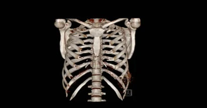
3D Rendering and Volume Calculation
In cases of complex fractures or organ tumors, DICOM data is used to create 3D Volume Renderings. Surgeons can literally "see" the 3D structure of an organ before they operate.
Cine Loops (Motion Imaging)
For Cardiac MRI or Ultrasound, DICOM supports "Cine" files—essentially high-definition videos of the beating heart or flowing blood, allowing for functional analysis (Ejection Fraction, etc.).
6. Common Misconceptions: The "CD" vs. The "Report"
Many patients in India receive a CD after their scan. That CD contains the DICOM files and a basic viewer.
- The Problem: Many home computers cannot run the heavy DICOM viewers.
- The Solution: Modern teleradiology platforms like ours now provide Zero-Footprint Viewers (ZFP). This allows doctors to view the full-quality DICOM images directly in a web browser or on a smartphone without losing quality.
7. The Future: DICOM and Artificial Intelligence (AI)
In 2026, AI algorithms are being integrated directly into the DICOM workflow.
- Auto-Labeling: AI can read the DICOM header and automatically label the vertebrae in a spine MRI, saving the radiologist time.
- Triage: If the AI detects a bleed in a brain CT DICOM file, it can move that case to the top of the reporting queue automatically.
8. Why Choosing a DICOM-Compliant Partner Matters
For diagnostic centers and hospitals, partnering with a firm that understands the intricacies of DICOM is vital for:
- Legal Protection: Ensuring all metadata is intact for medico-legal cases.
- Diagnostic Accuracy: Avoiding the "quality drop" associated with poor file handling.
- Long-term Storage: Keeping a digital "life-long" record of patient scans.