


 February 03, 2026 - BY Admin
MRI vs. CT Scan: A Clinical Deep-Dive into Choosing the Right Diagnostic Path
In the modern era of precision medicine, the diagnostic phase is the most critical juncture of patient care. As a radiologist, the question I am most frequently asked by both patients and referring physicians is: "Doctor, which is better—an MRI or a CT scan?"
The answer is rarely a simple "this or that." It is a nuanced decision based on the specific pathology we are hunting, the patient’s physiological state, and the urgency of the clinical scenario. At AITeleRadiology, we analyze thousands of scans monthly, and we have found that understanding the technical divergence between these two modalities is the key to an accurate diagnosis.
This comprehensive guide provides an expert-level comparison of Magnetic Resonance Imaging (MRI) and Computed Tomography (CT), ensuring you have the deep knowledge required to navigate Indian healthcare diagnostics in 2026.
1. The Fundamental Science: Radiation vs. Magnetism
To understand the clinical choice, we must first look at the physics under the hood.

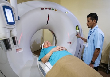
Computed Tomography (CT): The Power of X-Rays
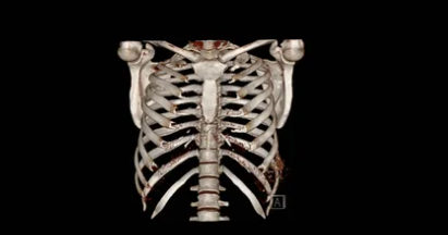
A CT scan is essentially a sophisticated evolution of the traditional X-ray. It uses a rotating gantry equipped with an X-ray source and electronic detectors. As the gantry spins around the patient, it captures multiple "slices" of the body. These slices are reconstructed to create a 3D volume.
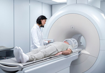
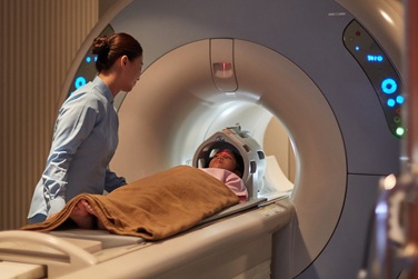
Magnetic Resonance Imaging (MRI): The Symphony of Protons
MRI is fundamentally different. It uses no radiation. Instead, it relies on a powerful magnetic field (usually 1.5T or 3.0T) and radiofrequency (RF) pulses to manipulate hydrogen protons in your body. When the energy is released, it is transformed into an image.
2. When Seconds Count: The Emergency Room Perspective
In clinical emergencies, the speed of acquisition is often the deciding factor.
Why CT Wins in Trauma
In the event of a road accident or a suspected brain hemorrhage, time is tissue. A CT scan of the head or abdomen takes less than 30 to 60 seconds. It is exceptionally sensitive to:
- Acute Bleeding: CT is the gold standard for identifying a subarachnoid or intracranial hemorrhage immediately.
- Bone Fractures: CT provides a detailed 3D map of complex bone breaks.
When MRI Takes the Lead in Emergencies
The only major emergency where MRI is superior is an Acute Ischemic Stroke. Using a sequence called DWI (Diffusion-Weighted Imaging), we can detect a stroke within minutes of onset, whereas a CT might appear normal for the first 6 to 12 hours.
3. Neuro-Imaging: Mapping the Human Brain
When we move into chronic conditions, the complexity of the brain requires the "high-resolution" lens of an MRI.
The Superiority of MRI for the CNS
The Central Nervous System (CNS) is shielded by bone. While CT can see the bone, MRI can "see through" it to the delicate structures within.
- Multiple Sclerosis (MS): MRI is the only way to visualize demyelinating plaques.
- Tumor Grading: Using MR Spectroscopy (MRS), we can analyze chemical metabolites inside a tumor.
- Dementia and Alzheimer’s: MRI can measure hippocampus volume to track atrophy.
4. Musculoskeletal (MSK) Health: Ligaments vs. Bones
For the orthopedic community in India, choosing between MRI and CT depends on what exactly is broken.
The MRI Domain (Soft Tissue)
If you have a sports injury, MRI is your best friend. It is the only modality that clearly visualizes:
- Ligament Tears: ACL, PCL, MCL in the knee.
- Cartilage Damage: Meniscal tears or labral tears.
- Nerve Impingement: Sciatica investigations.
The CT Domain (Complex Bone Work)
If a surgeon is planning a joint replacement or treating a complex "shattered" fracture, they will request a CT. The ability of CT to provide 3D reconstructions allows surgeons to "practice" the surgery digitally.
5. Thoracic and Abdominal Imaging
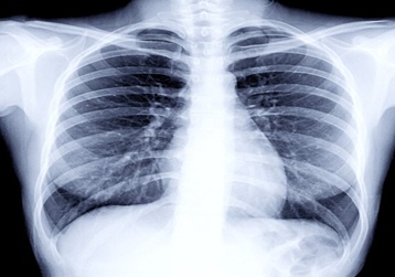
CT: The Guardian of the Lungs
The lungs are filled with air, which is a "black hole" for MRI. Therefore, CT is the undisputed king of chest imaging for conditions like lung cancer, pneumonia (ILD), or Pulmonary Embolism (CT Angiography).
MRI: The Expert of the Liver and Pelvis
MRI is used for deeper investigations like MRCP (looking at bile ducts without endoscopy) or detailed pelvic imaging for uterine fibroids and prostate cancer.
6. Patient Safety, Contraindications, and Comfort
As a healthcare provider, my first duty is Primum non nocere (First, do no harm).
CT Safety Concerns
- Radiation: Not suitable for pregnant women.
- Contrast: Iodinated dye can be hard on kidneys; Serum Creatinine check is mandatory.
MRI Safety Concerns
- The Magnet: Patients with pacemakers or metallic implants cannot enter.
- Claustrophobia: The narrow tunnel can be difficult for some; Wide-Bore machines are an alternative.
- Noise: Earplugs are mandatory due to high decibel levels.
7. The Economics: Cost vs. Value in India
In the Indian healthcare market, cost is a reality.
| Feature | CT Scan | MRI Scan |
|---|---|---|
| Approx. Cost (INR) | ₹2,000 - ₹7,000 | ₹6,000 - ₹15,000 |
| Availability | Widely available (Tier 2/3 cities) | Tier 1/2 cities, specialized centers |
However, the "value" of an MRI often outweighs the cost if it prevents an unnecessary surgery or a misdiagnosis.
8. The Role of Teleradiology in Accuracy
Whether you get an MRI or a CT, the machine only does 50% of the work. The other 50% is the Radiologist’s Interpretation. This is where aiteleradiology.in provides a global advantage.
A scan taken in a rural diagnostic center can be uploaded to our cloud and read by a sub-specialist radiologist in real-time, ensuring complex MRI sequences are read by experts and emergency CTs are reported in under 2 hours.